4 thoughts on “The Medial Prefrontal Cortex-Basolateral Amygdala Connection”
Hi Natalie,
I’m a bit confused because the title of this poster and the image that is displayed is for work conducted in Prof Blaise’s lab, but then when I click on the poster to view it in more detail what comes up is work you did with Dr. Nakanishi. But since the poster from Prof. Blaise’s lab is the one that is relevant for Neuroscience, I’ll ask questions about that one:
(1) How can you be certain that your recording electrode actually was successfully placed in the BLA? This is a relatively small brain area near the ventral surface of the brain, and you are approaching it from the dorsal surface. Obviously you have coordinates that you follow, but there are a lot of sources of variability in this kind of surgery, each of which could ultimately impact whether the electrode actually makes it to the targeted structure, or not.
(2) In the introduction it says the mPFC-BLA pathway does not show LTP, but then it also says that acute stress promotes LTP in this pathway. Please clarify.
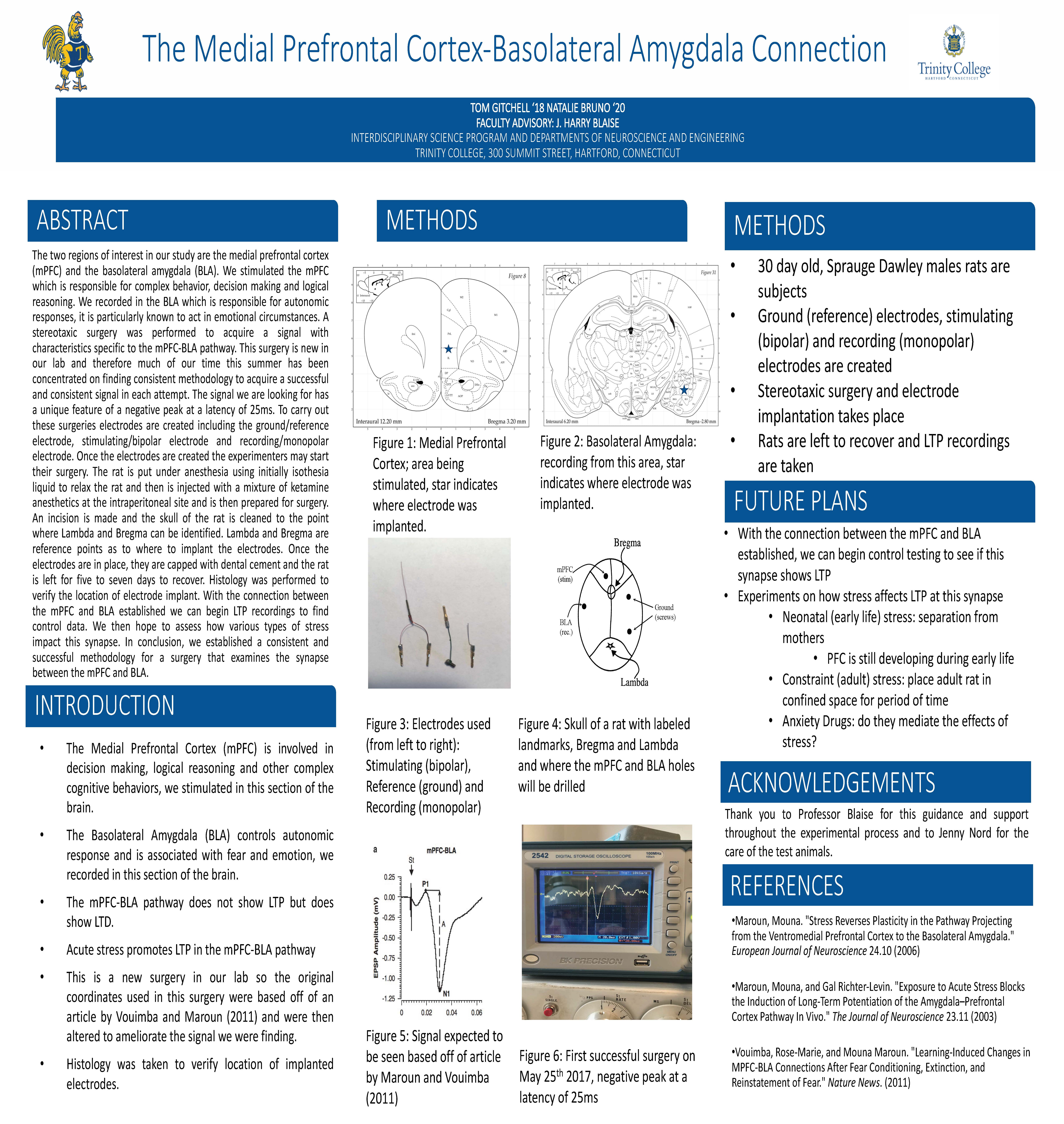
1. The area that was being targeted was extremely small. We spent the majority of summer assessing and reassessing our target regions within the brain by conducting many surgeries and adjusting as we went. We started our process by using the coordination points outlined in the 2011 article by Dr. Maroun and Vouimba. These original coordination points were not working for us to get the signal, it would either be very weak or come in and out. However, with practice, time and lots of literature research we created unique coordination points that worked very reliably for our surgeries. We suspected that the age and size of the rats may have been a variable as to why certain coordinations were working in some labs and not in others. We also found that adjusting the creation methods of the electrodes to make the recording wires shorter led to a more accurate signal from such a small target area. We were able to verify that we had the correct location in a couple of ways. Firstly, the signal that is recorded in the BLA-mPFC is very specific, so when we made the adjustments to the coordination points we could verify that the electrode was placed in the correct position. Secondly, while this was not represented visually on the poster we were in the process of moving forward with histology to verify the location of the electrodes in the brain. We conducted whole animal perfusion fixation and were allowing the brains to remain in a formaldehyde bath to fix the brain tissue for a longer amount of time before making slides. I was not able to complete the histology myself but this was our plan to verify the location of the implanted electrodes.
2. From the sample size (n=5) we used we did not see any significant changes in LTP from our latency testing on freely moving animals. The literature did indicate that acute stress does promote LTP in this pathway in rats under anesthesia but we could not find any data specifically pertaining to freely moving rats. Potentially there may be different effects of acute stress on mice who have more recently had surgery and whose latency testing was being conducted while they were still restrained and under anesthesia compared to the freely moving animals that we were interested in. To see a correlation of induced LTP in this region as outlined in the literature we should continue to increase sample size to verify a significant change in freely moving rats. The method of induced acute neonatal stress that was being used in this study was maternal separation. This was a new technique of inducing stress in our lab. To determine if there were changes to be seen in this pathway, specifically induced by acute neonatal stress, there may have been other more effective methods of inducing acute neonatal stress, for example, using restraint stress. We could verify the efficacy of the maternal separation protocol used by our lab to induce acute stress by assessing other pathways as well that have been shown to be influenced by acute neonatal stress. I would make sure to clarify this point of distinction in further editions of the poster.

Hi Natalie,
I’m a bit confused because the title of this poster and the image that is displayed is for work conducted in Prof Blaise’s lab, but then when I click on the poster to view it in more detail what comes up is work you did with Dr. Nakanishi. But since the poster from Prof. Blaise’s lab is the one that is relevant for Neuroscience, I’ll ask questions about that one:
(1) How can you be certain that your recording electrode actually was successfully placed in the BLA? This is a relatively small brain area near the ventral surface of the brain, and you are approaching it from the dorsal surface. Obviously you have coordinates that you follow, but there are a lot of sources of variability in this kind of surgery, each of which could ultimately impact whether the electrode actually makes it to the targeted structure, or not.
(2) In the introduction it says the mPFC-BLA pathway does not show LTP, but then it also says that acute stress promotes LTP in this pathway. Please clarify.
Best,
Prof. Martinez
Sorry, the poster confusion is my fault. I have fixed the pdf
Professor Martinez,
Thank you for your questions.
1. The area that was being targeted was extremely small. We spent the majority of summer assessing and reassessing our target regions within the brain by conducting many surgeries and adjusting as we went. We started our process by using the coordination points outlined in the 2011 article by Dr. Maroun and Vouimba. These original coordination points were not working for us to get the signal, it would either be very weak or come in and out. However, with practice, time and lots of literature research we created unique coordination points that worked very reliably for our surgeries. We suspected that the age and size of the rats may have been a variable as to why certain coordinations were working in some labs and not in others. We also found that adjusting the creation methods of the electrodes to make the recording wires shorter led to a more accurate signal from such a small target area. We were able to verify that we had the correct location in a couple of ways. Firstly, the signal that is recorded in the BLA-mPFC is very specific, so when we made the adjustments to the coordination points we could verify that the electrode was placed in the correct position. Secondly, while this was not represented visually on the poster we were in the process of moving forward with histology to verify the location of the electrodes in the brain. We conducted whole animal perfusion fixation and were allowing the brains to remain in a formaldehyde bath to fix the brain tissue for a longer amount of time before making slides. I was not able to complete the histology myself but this was our plan to verify the location of the implanted electrodes.
2. From the sample size (n=5) we used we did not see any significant changes in LTP from our latency testing on freely moving animals. The literature did indicate that acute stress does promote LTP in this pathway in rats under anesthesia but we could not find any data specifically pertaining to freely moving rats. Potentially there may be different effects of acute stress on mice who have more recently had surgery and whose latency testing was being conducted while they were still restrained and under anesthesia compared to the freely moving animals that we were interested in. To see a correlation of induced LTP in this region as outlined in the literature we should continue to increase sample size to verify a significant change in freely moving rats. The method of induced acute neonatal stress that was being used in this study was maternal separation. This was a new technique of inducing stress in our lab. To determine if there were changes to be seen in this pathway, specifically induced by acute neonatal stress, there may have been other more effective methods of inducing acute neonatal stress, for example, using restraint stress. We could verify the efficacy of the maternal separation protocol used by our lab to induce acute stress by assessing other pathways as well that have been shown to be influenced by acute neonatal stress. I would make sure to clarify this point of distinction in further editions of the poster.
Thanks,
Natalie
Hi Natalie,
Thank you for the clarifications.
Best,
Prof. Martinez